ISIC Skin Image Analysis Workshop
@ CVPR 2020
Hosted by the International Skin Imaging Collaboration (ISIC)
NEW (6/15/2020): Video of Opening Remarks posted
NEW (6/14/2020): Videos and Slides of Paper Presentations posted
NEW (6/14/2020): Videos of Invited Talks posted
NEW (6/14/2020): Open-Access Workshop Proceedings available
NEW (6/13/2020): CVPR Virtual Site launched
NEW (5/24/2020): SCHEDULE POSTED
NEW (3/9/2020): COVID-19 STATEMENT. DEADLINE EXTENSION TO MARCH 22nd, 11:59:59 EST
NOTICE: Paper Submission System Online
Skin is the largest organ of the human body, and is the first area of assessment performed by any clinical staff when a patient is seen, as it delivers numerous insights into a patient’s underlying health. For example, cardiac function, liver function, immune function, and physical injuries can be assessed by examining the skin. In addition, dermatologic complaints are also among the most prevalent in primary care [1]. Images of the skin are the most easily captured form of medical image in healthcare, and the domain shares qualities to other standard computer vision datasets, serving as a natural bridge between standard computer vision tasks and medical applications. However, significant and unique challenges still exist in this domain. For example, there is significant visual similarity across disease conditions, and in comparison to other medical imaging modalities, the skin is an organ which has no expected shape: varying genetics, disease states, imaging equipment, and environments can significantly change its appearance, making localization and classification in this domain unsolved tasks.
This workshop will serve as a venue to facilitate advancements and knowledge dissemination in the field of skin image analysis, raising awareness and interest for these socially valuable tasks. Invited speakers include (confirmed) major influencers in computer vision and skin imaging, and authors of accepted papers.
Topics of manuscript submissions may include:
- Computer Vision Applied to Dermatology and Primary Care
- Skin Analysis from Dermoscopic Images
- Skin Analysis from Consumer-Grade Photographs
- Skin Analysis from Video
- Skin Analysis from Total-Body Photography and 3D Skin Reconstructions
- Skin Analysis from Confocal Microscopy
- Skin Analysis from Optical Coherence Tomography (OCT)
- Skin Analysis from Histopathological Images
- Skin Analysis from Multi-Modal Data Sources
- Interpretable or Explainable AI Related to Skin Image Analysis
- Algorithms to Mitigate Dataset Imbalance
- Uncertainty Estimation Related to Skin Image Analysis
- Application Workflows for Skin Image Analysis
- Robustness to Bias from Clinical and User-Originating Photography
- Few-Shot Learning in Skin Images
- Best Paper Award: 4,000 USD
- Honorable Mention Award: 2,000 USD
In order to submit a paper, please refer to the following:
Paper Submissions System Link Available *NOW*!
(Author Information and Template)
[1] Lowell BA, et al. “Dermatology in primary care: Prevalence and patient disposition" In: Journal of the American Academy of Dermatology (JAAD), vol. 45, no. 2. 2001.
Invited Speakers
This workshop will feature several prominent names in the field of skin image analysis, including:
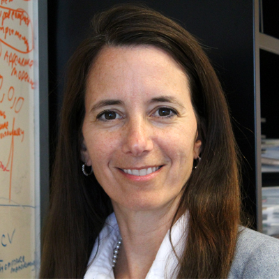 |
Dr. Dana is a Full Professor in the Department of Electrical and Computer Engineering at Rutgers University. She is also a member of the graduate faculty of Rutgers Computer Science Department. Her work on skin image analysis has included confocal microscopy analysis, skin texture quantification, and microbiome assessment, published in top venues such as CVPR and ICPR.. |
 |
Dr. Lee is an associate professor of Dermatology and Skin Science and a senior scientist of BC Cancer Research Centre. He is one of the earliest researchers in dermoscopy image analysis, and inventor of the DullRazor algorithm, which is used for automatic hair removal. |
 |
Doug Canfield is CEO of Canfield Scientific, one of the largest developers and manufacturers of dermatology imaging equipment, including dermatoscopes, facial imaging, total body imaging, and more. Canfield researches and develops automated analysis systems for their products using machine learning and artificial intelligence. |
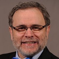 |
Dr. Siegel is a Clinical Professor of Dermatology at SUNY Downstate. He is a former President of the American Academy of Dermatology (AAD) and a member of the Scientific Board of SkinVision. |
Important Dates
| March 22nd, 2020: | Workshop Paper Submission Deadline: 11:59 PM EST |
| April 13th, 2020: | Author final notification |
| April 20th, 2020: | Camera-Ready Deadline |
| June 15, 2020: | Workshop @ CVPR 2020 Virtual |
Paper Submission
For paper submissions, CVPR manuscript rules are followed. All papers will be indexed in CVF and IEEE Xplore, along with other CVPR 2020 papers.
Research Datasets for Skin Image Analysis
- ISIC 2018 / ISIC 2019: According to the American Cancer Society, skin cancer is the most common form of cancer. While amenable to early detection by direct inspection, visual similarity with benign lesions makes the task difficult. Dermoscopic imaging was introduced to better visualize key details in skin lesions to improve diagnostic accuracy. The International Skin Imaging Collaboration (ISIC) has organized the world's largest repository of dermoscopic images of skin (over 23,000 images), to support research and development of methods for segmentation, clinical attribute detection, and disease classification. These datasets are snapshots used for the 2018 and 2019 challenges. See also: Challenge Summary / Data Description
- SD-198: 6,584 consumer grade photographs of skin affected by 198 disease states. See also: Paper Reference / Author Website
- Derm7pt: Over 2,000 dermoscopic and clinical images of skin lesions, with 7-point checklist criteria and disease diagnosis annotated.
Program Schedule
Location: Monday, June 15th, 7:45-13:00 PDT, WWW
| 7:45: | Opening Remarks (M. Emre Celebi) [Video] |
| 8:00: | Invited Talk 1: Computational Skin Texture (Kristin Dana) [Video] |
| 8:30: | Invited Talk 2: How to Advance Automated Computer-Aided Diagnostic System for Melanoma Detection (Tim K. Lee) [Video] |
| 9:00: | Invited Talk 3: 3D Total Body Photography and Change Detection (Doug Canfield) [Video] |
| 9:30: | Invited Talk 4: SkinVision: 21st Century Melanoma Screening with a Real Medical Device (Daniel Mark Siegel) [Video] |
| 10:00: |
Accepted Paper Presentation Session [Workshop Proceedings]
Chairs: M. Emre Celebi, Noel C. F. Codella, Marc Combalia, Kristin Dana, Allan Halpern, Philipp Tschandl
|
| 12:00: | Q&A Session (Live via Zoom) |
| 12:30: | Closing Remarks (Kristin Dana) |
Organizers
Sponsors:
Workshop Organizers:
- M. Emre Celebi Ph.D. (University of Central Arkansas, Arkansas, USA)
- Noel C. F. Codella Ph.D. (IBM Research, New York, USA)Twitter
- Marc Combalia M.Sc. (Hospital Clínic of Barcelona, Barcelona, Spain)
- Kristin Dana Ph.D. (Rutgers University, New Jersey, USA)
- Allan Halpern M.D. (Memorial Sloan Kettering Cancer Center, New York, USA)
- Philipp Tschandl M.D. Ph.D. (Medical University of Vienna, Vienna, Austria)
Steering Committee:
- Rogerio Feris Ph.D. (IBM Research, New York, USA)
- Anthony Hoogs Ph.D. (Kitware, New York, USA)
- John R. Smith Ph.D. (IBM Research, New York, USA)
- Harald Kittler M.D. (Medical University of Vienna)